

挑戦者の自己紹介

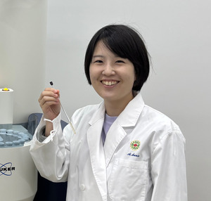
Annalisa Calò
My career as a scientist started when I was 25 years old, after a university degree … in Law! I knew I wanted to be a scientist, I loved chemistry and physics, but I was scared because of my non-scientific background. When I started my degree at the University of Bologna, my attention was caught by the word “nanotechnology”, which is the science of studying very small objects and single molecules, which are 50 000 times thinner than a human hair. I dedicated my PhD to learning the atomic force microscope (AFM) that allows touching and seeing these objects using a very small tip, like a nano-finger. After the PhD, I visited different research centers in Europe (Institute for Bioengineering of Catalonia IBEC) and USA (Advanced Science Research Center, ASRC CUNY) to learn other applications of the AFM. For example, I used it as a nanometric pen to write tiny electric circuits for next generation electronics. During 2018-2019, I worked in the Memorial Sloan Kettering Cancer Center (MSKCC) as a research expert in AFM. This experience was very fulfilling and helped me learn from biologists how they design their experiments and also envision new research paths where AFM can be a useful addition to biomedical research.
What is your research vision?
During my trajectory I was impressed by the capability of the atomic force microscope (AFM) to observe the nano-world. With it, it is possible to obtain images of very small objects, from viruses to single DNA molecules, without the need to treat them in a special way for the observation, like using vacuum or staining agents. I have even been able to recognize a few water molecules that absorb as thin layers on surfaces when humidity rises. The versatility of the AFM also allows not only seeing, but also distinguishing nanomaterials based on their properties, like the mechanical rigidity, electric charges or biochemical composition. When I worked in the hospital, I realized that living beings are also made up of nanomaterials. Thus, the capability of AFM to touch and probe nanomaterials can have enormous potential in medicine, because diseases are nothing but an expression of alteration of various proteins, ionic charges etc. in cells, tissues and organs. If cells, tissues, proteins are also materials, can the sensitive touch of AFM be used to watch and distinguish them? Why not use the AFM outcomes as markers of the wellness or malaise of a cell? In this way it can be employed to understand diseases or classify illnesses based on the effect that they induce on the functionality of cells and tissues. Currently, there is no equivalent microscope that allows measuring biophysical properties while watching, with such precision to resolve differences across nanometric areas of the sample without the need of biomarkers. We just need to use gentle forces to touch our sample and record the response! I strongly believe that my experience in advanced AFM research and in a medical setting will ensure successful accomplishment of my vision to bring the two worlds of x and y come closer.The final aim is to use the AFM as a resource to improve health and help society.

How do you realize your vision?
In order to integrate the AFM in the workflow of the clinical practice, I will use my established partnerships with hospitals and research centers connected to hospitals. This collaboration is aimed to tackle open problems where AFM measurements can be a valuable addition, to increase our knowledge on diseases or enlighten sample characteristics that cannot be resolved using other techniques (biochemical assays, histology, optical microscopy) due to their limitations. Also, new routes will be found where AFM can make biomedical testing less invasive, for example because it uses just small amounts of liquids, obtained from blood/urine samples, avoiding biopsies and surgical procedures. Partnership with hospitals is also necessary to obtain cells and tissues from patients, which are much more meaningful for disease investigation than commercially available samples, like cell lines. In order to transition AFM from the basic research in nanotechnology to the routine characterization of cells and tissues, a methodological change needs to be accomplished to have results available within the time of standard biomedical tests. Also, biological samples are often milli- to micrometer-sized, thus much larger than what AFM normally tests. So far AFM has been used to obtain images of nanometer areas with high pixel number; one such images usually takes 5-10 minutes. This throughput is not adequate for biological studies, where many cells and tissue locations need to be tested for the results to be statistically significant. Also, samples from many patients need to be analyzed. My approach will be focused on the development of protocols that allow for measurements with AFM to be performed in affordable timescales. Also, I will initiate collaborations to develop algorithms for data analysis that are based on the fast processing of big data in an unbiased way, instead of analyzing images one by one, as it is customary in AFM microscopy.

What is the research theme of this crowdfunding project?
The objective of this research project is to establish a protocol for the AFM imaging and analysis of big areas of biological samples, with millimeter size. This length scale is 5 orders of magnitude bigger than the scales which are normally tested by AFM. The protocol can be later on slightly adjusted depending on the specific sample, but a general frame will be provided as output in this project. As model samples, cancer cells with distinct phenotypes will be tested, in order to demonstrate the sensitivity of the AFM in distinguishing them as for the mechanical properties and demonstrate the protocol affordability as for time and robustness of the results. Fibroblast cells associated with different lung cancers (adenocarcinoma and squamous cell carcinoma), provided from the Clinic Hospital of Barcelona, will be tested and compared. It is important to be able to distinguish these two cell phenotypes, as drugs for one type of cancer may not work or can be harmful in the presence of the other cancer type. Mechanical mapping by AFM will be performed using colloidal probes and the measurement parameters (in particular, the pixel time and the travel distance to touch the sample) will be optimized in order to be able to test 100x100 micrometer square areas of the sample in less than 1 minute. Each patient dataset will contain in the order of tens of thousands stiffness data points that come from the observation of 10-20 cells. This data will be analyzed using commercial software for AFM data analysis integrated with custom-made scripts to make the analysis faster, more efficient and less manual. Also, a machine learning algorithm will be implemented as a new approach for automated and unbiased data analysis; results will be compared to those obtained with standard processing of AFM data.

Request for Research Funding Support
I think that a crowdfunding platform is a nice way to connect with people and make them aware of the research going on in different fields, some of them important for our future and well-being. Sometimes people feel science is difficult to understand and far from their everyday life and problems. Being able to explain it with simple terms can change the way people see it and luckily it can constitute a motivation for supporting science. I believe that positive feedback from society can also increase the motivation and passion of scientists towards their job, because they feel it is useful and appreciated by others. If I reach my crowdfunding objective, I will use the supported funds to upgrade the AFM system of my institute, in order to perform automated measurements with minimal manual operation and to purchase a license for data analysis. I will also use funds to purchase dedicated AFM probes for testing cells/tissues, chemicals, and to pay hospital and cleanroom facilities for sample preparation. Expenses for paper publication and conference participation will also be part of my expenditures. Once the measurement and analysis are established, I will be able to use it to create databases that include data of biophysical properties of cells and tissues for many patients and different illnesses. These biophysical data nowadays are not available in routine screenings and they can be used in the future to classify the gravity of a disease, to diagnose a disease where available techniques fail or to test the efficiency of a therapeutic plan from its impact on cell stiffness or charge. AFM results can in the future become a line in our blood test to check our wellness!
推薦者コメント
Dr. Prof. Elisa Riedo
I feel very lucky that Dr. Calò decided to move to NYC and work as postdoc in my group in 2016. I can say that she has been one of the best researchers that I have ever had (I have supervised about thirty students and postdocs). She has great passion and enthusiasm for science, and she is very meticulous and patient. As far as I know, the people who have been doing research with her all enjoy work and discussions with her. In addition, she learned what she could to sharpen her skills, increase the knowledge she needed, and widen her views in chemistry and physics.
Dr. Edoardo Albisetti
I have met Dr. Calò at the Advanced Science Research Center of the City University of New York (ASRC-CUNY), where we both were postdocs. We have been involved in the same project, and I enjoyed working with her. Besides her great talent as a scientist, she is an extraordinary individual at the human level. It is a pleasure to work in her company, and she has a great sense of humor, compassion, and resilience. She is always willing to help her colleagues, and she was indeed beloved by everybody. I still remember how many scientific discussions we have had and how many weekends we spent in the lab.

Dr. Sylvie Deborde
I enthusiastically recommend Dr. Annalisa Calò as a bright and tenacious scientist. She has been working in the imaging facility of my Institute, promoting AFM based research, training, managing and assisting facility users, maintaining the instruments at their optimum performance, setting up new instruments and measurements, developing facility user policies, working with administration and staff scientists of the facilities as a team. Very importantly, we established a close scientific partnership, she helped me with my project and we could get very good results together. Now that she is not in MSK anymore I am missing her.
研究計画
| 時期 | 計画 |
|---|---|
May-June 2022 |
Crowdfunding period |
July 2022 |
Development of the measurement protocol |
January 2023 |
Data analysis |
September 2023 |
Paper writing, conference presentation |
リターンの説明
We will send you a thank you message via email.
Thank you message
14人のサポーターが支援しています (数量制限なし)

Listing of supporter names in the research report / Thank you message
23人のサポーターが支援しています (数量制限なし)

Online Science Cafe Participation / Listing of supporter names in the research report / Thank you message
21人のサポーターが支援しています (数量制限なし)
Listing of supporter names in the paper / Online Science Cafe Participation / Listing of supporter names in the research report / Thank you message
8人のサポーターが支援しています (数量制限なし)

Individual discussion participation / Listing of supporter names in the paper / Online Science Cafe Participation / Listing of supporter names in the research report / Thank you message
0人のサポーターが支援しています (数量制限なし)

Listing of supporter names on Laboratory Website / Individual discussion participation / Listing of supporter names in the paper / Online Science Cafe Participation / Listing of supporter names in the research report / Thank you message
1人のサポーターが支援しています (数量制限なし)
当サイトは SSL 暗号化通信に対応しております。入力した情報は安全に送信されます。
Thank you message

14
人
が支援しています。
(数量制限なし)
Listing of supporter names in the research report 他

23
人
が支援しています。
(数量制限なし)
Online Science Cafe Participation 他

21
人
が支援しています。
(数量制限なし)
Listing of supporter names in the paper 他

8
人
が支援しています。
(数量制限なし)
Individual discussion participation 他

0
人
が支援しています。
(数量制限なし)
Listing of supporter names on Laboratory Website 他

1
人
が支援しています。
(数量制限なし)


